Uncovering the Mystery of Knee Pain: Exploring Why Certain Joint Deformities Cause Pain

Advanced imaging methods assist in distinguishing between symptomatic and asymptomatic discoid lateral meniscus (DLM) cases, with those showing symptoms often possessing increased meniscal coverage and height. This study gives insight into surgical planning and the reason for the development of symptoms in certain DLM patients.
Even with various high-end imaging technologies available to medical professionals for diagnosing tissue and joint anomalies noninvasively and with excellent accuracy, the conundrum remains: why do specific joint deformations cause symptoms in some individuals, while others remain symptom-free?
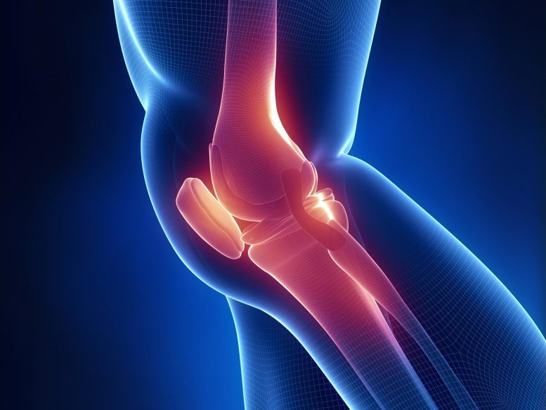
The knee joint is cushioned by a cartilage piece known as the meniscus, situated between the shin bone (or tibia) and the thigh bone (femur). A congenital morphological variation known as the DLM affects some individuals, causing a thickened meniscus on the knee's lateral (outer) side. Instead of the normal crescent shape, DLM deformations cause the lateral meniscus to take a circular shape, thus making the cartilage thicker and more susceptible to tearing. This results in symptoms such as knee pain and locking in some individuals, necessitating surgical intervention.
To determine the distinguishable factors between symptomatic and asymptomatic DLM cases, a research group led by Dr. Kazuya Nishino from Osaka Metropolitan University's Graduate School of Medicine carried out comprehensive studies. The study analyzed 61 knees undergoing surgery due to DLM without dislocation (symptomatic group) and 35 knees with DLM detected through MRI but without symptoms (asymptomatic group). The researchers calculated percentages of the meniscus in both coronal and sagittal sections and also measured meniscal height at its thinnest and thickest areas.
The symptomatic group had higher meniscal coverage in both coronal and sagittal planes and greater meniscal height when compared to the asymptomatic group. Picture Courtesy: Kazuya Nishino, Osaka Metropolitan University.
These findings highlight the significance of preoperative imaging in determining levels of tissue resection or removal required for symptomatic DLM patients. "Surgical decision-making and planning based on these characteristics is expected to aid in medical treatment. Future studies will focus on pre-and post-surgery meniscal shape changes in three dimensions," quoted Dr. Nishino.
Despite the most important finding being the diverse morphologies among symptomatic and asymptomatic DLM patients, MRI scans of symptomatic patients also revealed meniscal tears or signs of instability. The researchers propose that these morphological traits might be the cause of symptom development in cases of symptomatic DLM.


