Stunning Selections from Nikon’s Best Microscopy Images of 2024
With the snap of a camera shutter — and a handy microscope — what was once small can become grand.
Tap on the images below to enlarge
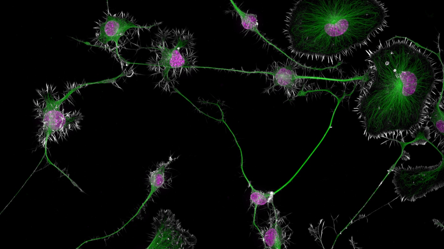
A close-up peek at mouse brain tumor cells has won first place in the 2024 Nikon Small World photomicrography competition. Neuroscientist Bruno Cisterna snapped the photo using a high-resolution microscopy technique as part of research to understand how neurodegenerative diseases such as Alzheimer’s and ALS develop.
The magenta blobs are the nuclei of each tumor cell, surrounded by actin proteins (white) that give the cells shape. In green are the rigid rods, or microtubules, that connect some cells and transport organelles within cells such as mitochondria, which generate much of a cell’s energy.
Earlier this year, Cisterna and colleagues included similar images in a study that found a protein called profilin 1, or PFN1, helps these microtubules function properly. Without enough PFN1, the team reported in the July Journal of Cell Biology, the cell’s microtubules transport more mitochondria faster around the cell, causing cells to die.
There’s a “delicate equilibrium” to how fast mitochondria can move around a cell and how many get transported, says Cisterna, of Augusta University in Georgia. Using images like the winning photo to track cellular structures can help reveal abnormalities that might be linked to cell death and neurodegeneration.
The tumor cells are one of 88 photos recognized October 17 in this year’s 50th annual contest. Here are a few other microscopic gems.
Sparks are flying between this entomology pin and a dismantled stick gas lighter.
Physicist and educator Marcel Clemens of Verona, Italy, used a wire to connect the lighter and pin, pulling the lighter’s trigger to set off sparks. Then, with long exposure times, he managed to capture electrical arcs traveling along the wire from the lighter to the pin. “The difficult thing was focus, and the only solution to this was trial and error,” Clemens says. “I have many hundreds of photos with out-of-focus sparks!”
Sponsor Message
The pink or purple glow comes from electrically charged atoms, or ions, that are losing or gaining electrons, Clemens says. But he’s less certain about where the blue emissions around the lighter come from. Nor does he know why there are orange trails coming off the top of the pin. But they could be from small particles of super-hot metal, he says, perhaps around 2,000° or 4,000° Celsius.
Photographer Paweł Błachowicz combined several dozen photos to create this “unearthly” closeup of a small crab spider’s eyes.
At true scale, the spider’s head is roughly 1 millimeter in size. Diaea dorsata males can grow to be up to 4 millimeters long and females up to 6 millimeters. Found in forests across Europe, these crab spiders can change color to blend in with their surroundings, although it takes several days.
At first glance the image might make some people think of a UFO, says Błachowicz, of Bedlno, Poland. For others, the close-up resembles a mutant Kermit the Frog.
These oval-shaped pairs might look like coffee beans. But they’re actually parasites.
Using a high-resolution microscope that uses lasers to image samples in three dimensions and a computational method to get a clear picture of immobile, physically expanded cells inside a gel, life scientist Kseniia Bondarenko snapped multiple photos of Toxoplasma gondii parasites dividing asexually inside a human skin cell. The parasites’ inner shells are colored magenta, the outer skeletons in yellow and the nuclei in blue.
Most people infected with the parasite — contracted from eating undercooked meat or contacting cat feces — don’t get symptoms. But those who do can have muscle aches or fever, and severe disease can cause damage to the brain and other organs. Once inside a cell, the parasites divide through a process called endodyogeny. As shown in the bottom left of the image, a mother cell is consumed as two daughter cells form. “The remnants of the mother ‘shell’ lie like a blanket wrapped around the two freshly emerged daughters,” says Bondarenko, of the University of Edinburgh.
Zoologist Sherif Abdallah of Tanta University in Egypt can find beauty in destruction.
Red palm weevils (Rhynchophorus ferrugineus) are among the most destructive palm pests in the world. “It uses its drill-like snout to bore into the heart of palm trees, leaving them hollow and ultimately destroying them,” says Abdallah, who is researching ways to combat the species. “What makes them even more dangerous is their ability to fly long distances, spreading infestations rapidly and turning entire groves into infested zones if not controlled.”
Abdallah used controlled lighting and stitched multiple images together to produce a snapshot of the insect’s head and highlight small details. With front antennae “curled forward like a boxer’s gloved fists,” it seems this red palm weevil is ready to put up a fight.
German photographer Daniel Knop loves to capture tiny details happening behind the scenes of natural phenomena. This striking series of photos of a swamp rose mallow (Hibiscus moscheutos) is no exception.
Knop placed a millimeter-sized H. moscheutos anther — the part of the flower that carries pollen — in front of a microscope and watched as the pod opened over a 40- to 50-minute span. In the folded state, minuscule balls of spiky pollen are barely visible. As the husk peels back, the pollen takes center stage.
It took hundreds of photos to capture each individual stage, Knop says. The resulting image gives viewers some insight into the development of living things and, he hopes, “make them curious to explore nature.”